
Have you been diagnosed with a brain injury? Or are you suffering from any of the following symptoms associated with traumatic brain injury?
- Mood swings
- Depression
- Memory loss or loss of cognitive function
- Speech or word recall difficulties
- Fatigue
- Loss of motor skills
- Sexual dysfunction
If you’ve suffered a head injury, it is also possible you may also be displaying very few outward physical indicators of your injury. Traumatic brain injuries are often called “the silent disability” – the symptoms are severe but often the visual indicators can be minimal.
“Mild” traumatic brain injuries – which make up 80% of the 1.5 million traumatic brain injuries suffered every year – are generally classified as a concussion, which is a loss of consciousness or confusion and disorientation lasting less than 30 minutes with a Glasgow coma scale of 13-15.
YOUR CASE AND TBI TECHNOLOGY
The correct diagnostic tools for traumatic brain injury technology help doctors understand the nature and extent of your injury and get you the treatment you require. If your case ends up going to court, these images can provide the type of scientific evidence that juries will find incredibly persuasive, especially when used to back up the other subjective complaints of the symptoms above.
Having an attorney with the knowledge of the right assistive technology to diagnose and treat traumatic brain injury is absolutely essential.
Your attorney should be able to:
- Understand what tools your doctors are using
- Be able to work with your medical team to get you the right assistive technology for diagnosis and treatment of your TBI
- Leverage this technology to make a powerful and persuasive case to the jury
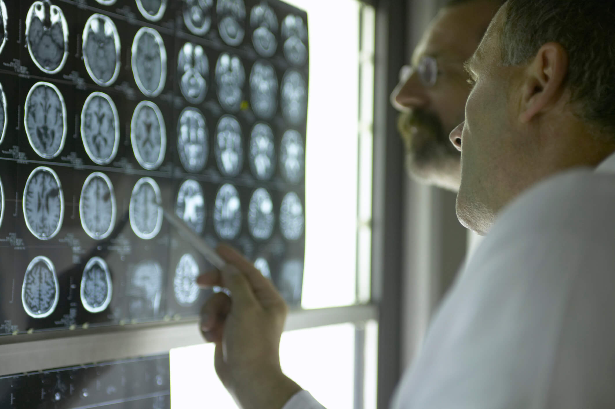
TYPES OF TRAUMATIC BRAIN INJURY TECHNOLOGY
CT Scan
- Effective in detecting bleeding in the brain (hematoma) or swelling in the brain (edema) that might require emergency treatment
- Limited in the ability to detect microscopic injuries to axons (nerve fibers) which transfer signals across the brain
- A big picture tool, but can frequently be completely normal for individuals suffering from TBI
MRI (Magnetic Resonance Imaging)
- A painless radiological scan that uses magnetic fields to develop images of your brain. There are no known side effects.
- Detects minute signs of injury such as minute bleeding (microhemorrhage), small areas of bruising (contusion) or scarring (gliosis)
- Even an MRI may not be able to detect microscopic injuries associated with brain injury
- When applied to mild traumatic brain injury, only about 10% of CT scan and 30% of standard MRI reveal abnormalities such as subdural or subarachnoid hemorrhage.
- Proven insensitive to subtle changes such as diffuse axonal shearing
Volumetric MRI
- A non-invasive brain imaging technique
- Used to measure volume and structure of specific regions of the brain
- Primarily used to measure atrophy of certain regions of the brain
- Brain atrophy means a loss of neurons, and the connections between them
- Atrophy can signal a decline or loss of cognitive ability, and while it can come naturally with age, a traumatic brain injury can accelerate the onset of atrophy or create permanent cognitive deficits.
Functional MRI (fMRI)
- Measures cerebral blood flow using the blood oxygen contrast
- Increased flow = increased neuron activity
- Used when a patient is performing a task versus at a resting state, to see if the portions of the brain associated with those tasks are firing correctly.
- Disruptions in activity have been found with patients suffering from mild traumatic brain injury, and increases in activity of certain areas of the brain are seen as mechanisms of compensation for other injured portions.
- Long term studies using fMRI show reduced activity of portions of the brain during memory tasks, making these scans useful tools to show the long-term effects of traumatic brain injuries.
Diffusion Tensor Imaging (DTI)
- An advanced MRI technique that measures diffusion properties of water molecules in the brain.
- Measures the manner and direction in which water molecules move throughout the microstructure of the brain
- The main imaging tool used to reveal diffuse axonal injury, it can reveal “white spots” on the brain where there is microscopic damage to myelin sheaths or axon membranes that otherwise would be undiscoverable.
- New measures have begun to increase sensitivity and specificity of the DTI, making it a more common tool. It is the most sensitive non-invasive method for detecting white matter changes as a result of mild TBI.
USING TBI TECHNOLOGY TO DEVELOP YOUR CASE
Traumatic brain injury technology plays a vital role in correctly diagnosing your injury and getting the care you require. The right brain-injury imaging allows the attorneys at Brain Injury Law of Seattle to assist you in your recovery process, help you get the treatment you need, and find the right doctors for you. Contact our traumatic brain injury lawyer today!
When it comes time to convince a jury of the nature and extent of your injuries, our lawyers know how to effectively present this technology to a jury by combining your personal story with scientific evidence to communicate exactly how the injury has changed your life.
Related Article:
Cutting Edge Technology That Can Help Prove Your Brain Injury
Most Common Sports Injuries
Road Rash Healing Process
Post-Concussion Syndrome
