Mild brain injuries have been called the “silent epidemic.”
This is because X-Ray and imaging technology has been trailing far behind brain science. So while doctors and brain injury attorneys have known to blame certain symptoms on brain injuries, there has never been a way to prove that a mild brain injury has taken place — until now.

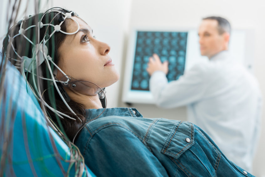
Vital procedure. Beautiful dark-haired woman lying on an examination table and undergoing electroencephalography while her doctor examining CT results
Traditionally, and even after the invention of CT and MRI scans, which are normal 90% of the time, the only way to really test for the invisible fingerprints of brain injury has been to submit the patient to a series of neuropsychological tests. These cognitive tests try to determine how and where cognitive deficits like memory, concentration, and thinking have been affected. A neurologist will then use the test results to infer what part of the brain has been injured.
Unfortunately, even with these tests, attorneys have been hard-pressed to convince a judge and jury that a brain injury has occurred.
This is because cognitive tests aren’t always considered objective proof of brain injury. The defense attorneys and their doctors in brain injury cases know this and have a long track record of manipulating neuropsychological test results to be interpreted against the patient. This is an easy task for the defense, as these tests always include a personality test section which allows them to infer that brain injury symptoms might be the result of depression or other mental conditions.
For reasons like these, the silent epidemic of mild-to-moderate brain injury has been allowed to ravage its victims unnoticed and remain susceptible to the insurance defense attorneys’ unfounded logic of “no objective proof = no brain injury.”
We intend to stop insurance defense attorneys from improperly labeling brain injury victims.
Here at Brain Injury Law of Seattle, our team of experts is using industry-leading assistive technology to break the silence and prove the existence of these formerly “invisible” traumatic brain injuries.
In this blog post, I will introduce you to the silent epidemic, the technology that protected it, and the new assistive technology we are using to fight it.

Page Contents
A BRIEF HISTORY OF THE SILENT EPIDEMIC AND THE TECHNOLOGY THAT PROTECTED IT
Let’s start our journey in the 1970s.
During this time there wasn’t any effective brain imaging technology for TBIs. The brain was a “black box”, so when someone suffered a head injury, the only real option available was for neuropsychologist to assemble the person’s symptoms through testing and then try to piece the puzzle together to determine if there was a brain injury.
This method of sleuthing out brain damage indirectly worked, in that doctors could only partially infer if someone did have a brain injury. Nonetheless, these techniques couldn’t determine which specific part of the brain had been damaged and couldn’t produce the kind of objective proof that easily compels a jury to side with the plaintiff.
Sadly, in the ‘70s, the only real way to locate brain injuries was by conducting an autopsy, which of course, couldn’t be done on a live person.
By the mid-‘80s, the CT scan had come on the scene and was starting to be used in America. While this was a massive step forward for brain technology, the CT scan could only be used to reveal significant brain injuries like brain bleeds and tumors. A CT couldn’t give enough detail for neurologists to look at the brain and say, “Yep! This is the part that is injured.”
So despite this new assistive technology, anything short of a tumor, brain bleed, lesion, or some other kind of mass in the brain, went unnoticed. Mild and moderate traumatic brain injuries continued to go unresolved because they couldn’t be identified.
Then in the early ‘90s, the MRI was developed. These machines were significantly better than the CT scan but were still unfortunately limited in their sensitivity.
MRIs do a better job exposing unnatural masses or significant changes to brain structure but don’t get granular enough to show microscopic brain injuries unless they are extensive. This means that the vast majority of brain injuries can’t be identified by MRI. For example, when someone has a concussion and gets an MRI scan to identify it, 90-95% of the time, the MRI will show that the brain is normal — it can’t detect the microscopic damage giving rise to the concussion.
Other tools like a Spect Scan became available, but once again, it did not provide convincing evidence of brain injury, and that brings us to today!
Up until very recently, the principal clinical tools available to detect brain injuries have been CT and MRI scans, and as explained above, both of these technologies often fail when it comes to providing objective proof of mild-to-moderate brain injury.
So why all this trouble? Why are concussions and mild brain injuries so difficult to identify?
To answer that, let’s take a look inside the skull to see what happens to your brain during an injury-inducing event.

WHAT IS A CONCUSSION?
A concussion can occur when the head experiences trauma. For example, if your head gets struck by something, or is thrown from side to side, or back and forth (e.g. whiplash).
An action like these causes the brain to smash against the inside of the skull. Imagine if you have ever had to hit the bottom of a jar with liquid in it so you can loosen the screw top lid, and you can feel the shock wave go through the jar and hit the lid with enough force to loosen the cap. This is the same thing that happens to the brain. This unnatural brain movement tears brain cells apart, which leads to the brain having trouble operating the way it was before.
Stated differently, it is similar to how if you drop your computer on the ground, then it might develop problems because certain connections and wires have been broken.
Now, you might be thinking, “If I hit the front of my head, that means the front of my brain was smashed. So that’s probably where my brain injuries are!”
Unfortunately, it’s not that simple.
You have to look at the brain as an exquisite electrical machine made up of complex and unique parts, kind of like a symphony. Your brain is an interconnected support system — affect one part of it and the whole system will be affected as well.
Because the brain functions systemically, a concussion tends to alter the balance within the entire brain, which is why someone might complain about foggy thinking, dizziness, and vision problems after striking their head.
WHY ARE CONCUSSIONS AND MILD BRAIN INJURIES SO SERIOUS?
Your brain is about the consistency of hard jello.
So you’ve got this tender mass confined in your bony skull and when you smack your head, yes, the point of impact is affected, but you’ve also got a shockwave that gets sent through the brain.
This shockwave disrupts connections along its path and causes an impact on the opposite side of the brain.
So there are three areas that are generally affected during a concussion:
- The point of impact
- The pathway of the shockwave
- The opposite side of the brain
Now, what are these connections that are disrupted by the impacts and shockwave? To understand this, you need to know a little bit about how brain cells function.
A brief introduction to brain cells and their function
The brain is made up of about 85-100 billion brain cells. Each one of these cells has three parts: the nucleus, the axon, and the dendrites. The axon and dendrites extend from the nucleus and can be quite long.
Every brain cell is reaching out and using its dendrites to attach itself to the cells around it. There are literally trillions of connections. And this is why the brain functions systemically — it’s all connected.
This is why when you have a thought, sensation, or feeling, this impulse is passed from brain cell to brain cell, getting passed along via the dendrites and processed systemically.
What happens when the brain gets concussed is the connections between the dendrites are torn, making it difficult to pass thoughts along brain cells through your brain.
To help you visualize this, imagine your brain is like the electric grid in a city, with power lines representing the dendrites. A concussion is like an earthquake, which goes through your city and knocks down some of the power lines. Suddenly, parts of your brain city are having trouble receiving power, resulting in flickering lights and blackouts in various parts of the city.
In medical terms, this earthquake and the dendrite-tearing process is called “diffuse axonal shearing.” So you’re literally seeing brain cells get torn apart on the microscopic level, resulting in memory loss, problems thinking, headaches, and a great host of other possible TBI symptoms.
Unfortunately, brain cells are so small, that damage like this cannot be picked up by CT scans and MRIs. This is why 95% percent of concussions go unnoticed by MRIs — the technology can’t see the torn connections between dendrites. It’s rather like a satellite with a cell phone camera trying to photograph the downed power lines following the earthquake from outer space you just can’t see them.
This leads the neurologist to say, “Well, nothing looks damaged.” But this doesn’t mean the person doesn’t have a brain injury.
In an attempt to compensate for ineffective assistive technology, psychologists developed neuropsychological testing to test for brain injury. Regrettably, what was meant to be a solution when we didn’t have good imaging turned out to be a bane — which brings us to…

NEUROPSYCHOLOGICAL TESTING: A LIMITED SOLUTION FOR IDENTIFYING BRAIN INJURIES
CT and MRI scans were certainly advanced imaging technologies, but they couldn’t detect the invisible.
Medicine then had to rely on neuropsychologists, who as stated above, created a series of cognitive tests that they hoped could detect where in the brain trauma had occurred.
The only way to assess what kind of damage was done in mild or moderate brain injury situations, was to sit the patient down and have them do neuropsychological testing.
Neuropsychological testing does have its uses, but the practice is, in my opinion, a very limited method used to detect brain injury.
What is neuropsychological testing?
Basically, neuropsychological testing is an indirect way of peering into the brain.
This is done via a series of cognitive tests. They sit you in a quiet room with no external stimulation and test you on all sorts of different tasks, like memory, word-finding, reasoning and cognitive recall.
Based on how a neuropsychologist scores these tests, they can then be passed on to a neurologist, who will infer whether or not you have received a brain injury and where in the brain the injury is. But even then, this is hard, because as I explained earlier, the brain functions as a system, so pinning the injury to a single part of the brain is sketchy work at best.
For decades, this kind of testing has been seen as the industry standard for mild-to-moderate brain injury. This is because more direct forms of brain testing, like CT and MRI, have been unable to detect injuries like diffuse axonal shearing on their own.
How neuropsychological testing can negatively affect your brain injury case
Neuropsychological testing has a lot of limitations. One of the worst is that when doing these tests, almost every neuropsychologist also gives the patient an MMPI (Minnesota Multiphasic Personality Inventory).
The MMPI is not a brain injury test. It’s a personality exam.
The MMPI isn’t designed to detect brain injuries, it was created to analyze someone who has potential mental health problems. Consequently, the inclusion of the MMPI means that the neuropsychologist is entertaining the possibility that a person’s brain injury symptoms are possibly due to a mental or behavioral disorder. The insurance industry loves this test because it allows them to hire neuropsychologists loyal to insurance companies to make the plaintiff’s character the issue, not their brain injury. In other words, they will claim the plaintiff is faking, malingering, or simply depressed as an alternative to the brain injury since a brain injury is not easily seen on MRI.
As you can imagine then, having the MMPI in a plaintiff’s medical record is basically like handing the defense a shiny, wrapped gift.
And the sad truth is that insurance defense attorneys get away with this because they know there is very little any objective evidence that can actually prove brain damage. This has been a historical problem for the legal community and is why the “silent epidemic” of mild-to-moderate brain injuries has gone on for so long in legal cases.
It doesn’t need to be this way though. As I’ll explain in a moment, there are now powerful assistive technologies available that have the ability to prove brain injury.
Cutting Edge Technology is Here.
As time has gone by we have seen more and more really good imaging techniques start to develop.
THE ASSISTIVE TECHNOLOGIES WE USE TO PROVE BRAIN INJURIES
Rather than doing only neuropsychological testing like many lawyers do, our approach is to get as much objective testing as possible so that we can prove that a brain injury has occurred.
This approach does three things:
- When we go to trial, the jury can see and appreciate that there is objective evidence.
- It takes away the standard MMPI excuse of the defense that our client is simply suffering from unrelated mental disorders.
- This also significantly increases the odds of a jury or judge finding that a brain injury has occurred.
There are a number of other objective tests that we use to our benefit. The rest of this blog post is dedicated to explaining what these tests do and how they provide objective proof of brain injury.
These are the tools we are using to see the invisible and end the “silent epidemic.”
Diffuse Tensor Imaging
In addition to the fMRI, there are some enhanced software packages called Diffuse Tensor Imaging (DTI) that can be used with some MRIs. This is an incredible form of imaging that takes advantage of the water protons that move around brain cell dendrites.
When diffuse axonal shearing occurs, it leaves behind microscopic scar tissue that prevents water protons from moving around the dendrites in the normal way. What DTI does is reveal white matter brain injuries by highlighting these areas where water protons can no longer flow normally around brain cells.
DTI testing creates beautiful and compelling images that we use in court to provide objective proof of a brain injury. This kind of testing is a well-accepted science — especially when supported by the symptoms of brain injury.
Arterial Spin-Labeling
Another form of objective testing is Arterial Spin-Labeling (ASL). This is a cutting edge piece of brain injury detection software.
The way it works is by putting the patient in a 3T MRI, which magnetizes the protons in the blood. Once the blood is magnetized, the ASL tracks the blood as it moves up through the carotid artery and into the brain. This data gets visualized and allows the doctor to see what parts of your gray matter have scarring and brain damage.
Brain Injury Law of Seattle, is one of the few law firms in the country that uses ASL testing to provide objective evidence that brain damage has occurred.
Electroencephalography
Electroencephalography (also known as QEEG brain mapping) is a form of brain testing where a bunch of electrodes are placed on a person’s head. These electrodes track the brainwaves that are moving around inside the brain.
While DTI and ASL measure the interior structure and “plumbing” within the brain, QEEG is rather like looking at the electrical functioning of the brain. It provides an overview of the brain and identifies what regions of the brain are producing weaker electrical brainwaves than should be generated by the brain.
QEEG brain mapping is another way our law firm provides objective proof of brain injury.
Volumetric Studies
Another method of objective testing that we do is called volumetric studies. These tests are used to measure loss in brain volume over time, which can be indicative of brain damage.
Here’s how volumetric studies work.
When you receive mild-to-moderate brain damage, it is common for this event to damage brain cells inside of your brain. This leads to scarring and reduction in blood flow to the damaged areas, which in turn leads to atrophy and shrinkage in those areas overtime.
This shrinkage is referred to as “volume loss.”
If you have an MRI taken shortly after the injury, and then you have another MRI done a year or two later, then a neurologist will be able to compare these two brain scans. This will enable them to determine the exact percentage of volume loss that has occurred.
What differentiates brain-injury volume loss from the natural shrinkage that results from aging?
If a person is just getting old, then you will see volume loss evenly distributed around the brain — uniform reduction like this is not typically due to trauma. On the other hand, if the volume loss is asymmetric, this means that brain injury has likely occurred. This science makes volumetric studies compelling evidence in a brain injury court case.
Related Articles
OBJECTIVE PROOF MAKES ALL THE DIFFERENCE
Here at Brain Injury Law of Seattle, we are driven by the hope of helping our clients recover from their injuries.
This is why we don’t settle for only neuropsychological testing. We don’t leave any room for the defense to claim that your symptoms are the result of “childhood problems” or “character flaws.”
When you partner with us, we make sure that your case is supported by compelling, objective proof of brain injury. Our team of experts who use the industry-leading assistive technology described in this blog post to break the “silence” and prove the existence of your brain injuries.
If you would like to hear more about how we are bringing the “silent epidemic” to an end, or would like to schedule a consultation, contact our office today.
Useful Resource
Car Accident Injury Lawyer


 Brain Injuries")